Your position:Home > Products > Obstetrics & Gynecology Equipments > Mammography X-ray System
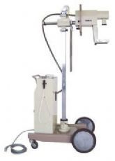
Features
It is used to diagnose early mamma pathological changes.
Using the Mo. target X-ray tube can show the pathological changes details.
A nipple areola cuticles fat galactophore canals glandular tissue connective tissue and blood vessels can be seen in the picture.
It has high correctness for distinguishing benign tumor and malignant tumor.
It can be moved to sickroom to photograph beside bed.
The unit can also be used to find foreign matters in human body to do nondestructive inspection of right metal and nonmetal materials.
|
Main character |
Shockproof, single focus and full-wave rectification |
|||
|
|
Current |
30mA |
||
|
Voltage |
34kVp |
|||
|
Time sec. |
2s |
|||
|
|
Voltage |
180-240V |
||
|
Frequency |
50Hz |
|||
|
Power |
Not lower than 1.3VA |
|||
|
Range of the timer |
0.4-2s |
|||
|
Film size |
127mm x 178mm |
|||
|
|
Vertical movement range |
630mm |
Adapt to different part of human |
|
|
|
|
|||
|
The Camera Head Mounting horizontal range |
160mm |
|||
|
Rotating angle of the module of the Camera Head Mounting and pole |
180° |
|||
 皖公网安备 34081102000223号 Copyright © 2017 Anjue Medical Equipment Co., Ltd. All rights reserved. 皖ICP备19019483号